Case #472 – July, 2018
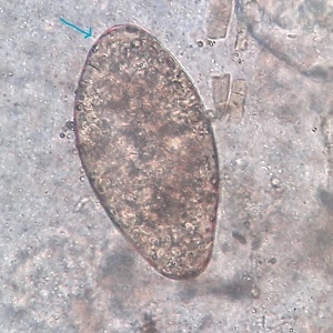
A 22 year old man diagnosed with gastroenteritis underwent subsequent gastrointestinal endoscopic examination. The gastroenterologist observed a worm which he extracted from the duodenum and sent to the laboratory for identification. Stool samples were also processed for ova and parasites (O&P) examination. Images of the worm and objects seen in a wet mount preparation from a formalin-ethyl acetate (FEA) concentration of the stool samples were captured by the laboratory technologist and sent to DPDx for identification. Travel history was not provided. The objects in Figures C – E measured 130 µm in length by 65 µm in width on average. The adult worm measured approximately 70 mm by 18 mm. What is your diagnosis? Based on what criteria?
This case and images were kindly provided by The Institute of Medicine, Tribhuvan University and Teaching Hospital, Nepal.

Figure A

Figure B

Figure C

Figure D

Figure E
Images presented in the dpdx case studies are from specimens submitted for diagnosis or archiving. On rare occasions, clinical histories given may be partly fictitious.
DPDx is an educational resource designed for health professionals and laboratory scientists. For an overview including prevention, control, and treatment visit www.cdc.gov/parasites/.
