Key points
- Anotia (an-NO-she-uh) and microtia (my-KRO- she-uh) are birth defects of a baby’s ear.
- Anotia/microtia affects how the baby's ear looks, but usually the parts of the ear inside the head are not affected.
- Researchers estimate that 1 in every 3,800 babies is born with anotia/microtia in the United States.
What it is
Anotia/microtia develops during the first few weeks of pregnancy. These defects can vary from being barely noticeable to being a significant problem with how the ear formed.
Usually, anotia/microtia only affects how the baby's ear looks. Sometimes, the parts of the ear inside the head are affected, and babies will have a narrow or missing ear canal.
Anotia/microtia can occur alone, with other birth defects, or as part of a syndrome.
Microtia
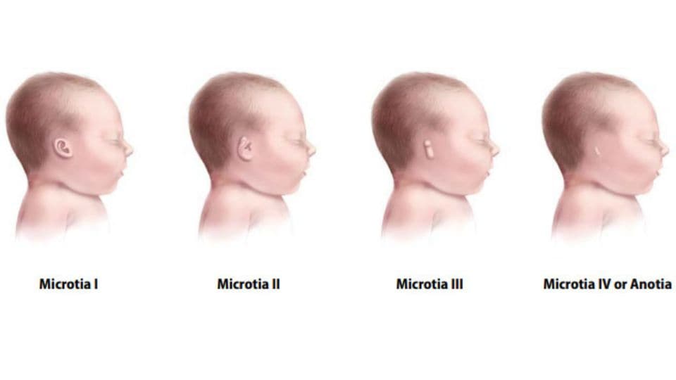
Microtia is when the external ear is small and not formed properly.
There are four types of microtia. Type 1 is the mildest form, where the ear has its normal shape, but is smaller than usual. Type 4 is the most severe type where all external ear structures are missing —anotia.
This condition can affect one or both ears. However, it is more common for babies to have only one affected ear.12
Anotia
Anotia is when the external ear (the part of the ear that can be seen) is missing completely.

Risk factors
The causes of anotia/microtia among most infants are unknown. Some known causes of anotia/microtia include:
- Certain changes in the baby's genes or chromosomes3
- Use of medicines such as isotretinoin (Accutane®) during pregnancy3
Research studies have also found some factors that might increase the risk of having a baby with anotia/microtia:
Diagnosis
Anotia/microtia is visible at birth. A healthcare provider can identify the problem by examining the baby. A CT or CAT scan (special x-ray test) of the baby’s ear can provide a detailed picture of the inner ear. This will help the provider see if the bones or other structures in the ear are affected. The provider should also perform a physical exam to look for any other birth defects that may be present.
Treatment
Treatment for babies with anotia/microtia depends on the severity of the condition. A healthcare provider or hearing specialist called an audiologist will test the baby's hearing to determine if there is any hearing loss. Hearing aids may be used to improve a child's hearing ability and to help with speech development. Hearing loss should be addressed, as even loss in one ear can affect child’s ability to develop communication, language, and social skills.6
Surgery is used to reconstruct the external ear. The timing of surgery depends on the severity of the defect and the child’s age. Surgery is usually performed between 4 and 10 years of age.
Children with anotia/microtia can develop normally and lead healthy lives. They may have issues with self-esteem if they are concerned with visible differences between themselves and other children. Parent-to-parent support groups can prove to be useful for new families of babies with anotia/microtia.
Resources
The views of these organizations are their own and do not reflect the official position of CDC.
Children's Craniofacial Association (CCA): CCA addresses the medical, financial, psychosocial, emotional, and educational concerns relating to craniofacial conditions.
The National Craniofacial Association (FACES): FACES is dedicated to assisting children and adults who have craniofacial disorders resulting from disease, accident, or birth.
- Shaw GM, Carmichael SL, Kaidarova Z, Harris JA. Epidemiologic characteristics of anotia and microtia in California, 1989-1997. Birth Defects Research (Part A): Clinical and Molecular Teratology. 2004;70:472-475.
- Canfield MA, Langlois PH, Nguyen LM, Scheuerle AE. Epidemiologic features and clinical subgroups of anotia/microtia in Texas. Birth Defects Research (Part A): Clinical and Molecular Teratology. 2009;85:905-913.
- Luquetti DV, Heike CL, Hing AV, Cunningham ML, Cox TC. Microtia: epidemiology and genetics. American Journal of Medical Genetics Part A. 2012 Jan;158(1):124-39.
- Tinker SC, Gilboa SM, Moore CA, Waller DK, Simeone RM, Kim SY, Jamieson DJ, Botto LD, Reefhuis J, Study NB. Specific birth defects in pregnancies of women with diabetes: National Birth Defects Prevention Study, 1997–2011. American Journal of Obstetrics and Gynecology. 2020 Feb 1;222(2):176-e1.
- Ma C, Shaw GM, Scheuerle AE, Canfield MA, Carmichael SL, and National Birth Defects Prevention Study. Association of microtia with maternal nutrition. Birth Defects Research (Part A): Clinical and Molecular Teratology. 2012;(94):1026-1032.
- Kuppler K, Lewis M, Evans AK. A review of unilateral hearing loss and academic performance: Is it time to reassess traditional dogmata? International Journal of Pediatrics and Otorhinolaryngology. 2013;77: 617-622.
- Stallings EB, Isenburg JL,Mai CT, et al. Population-based birth defects data in the United States, 2011–2015: A focus on eye and eardefects.Birth Defects Research. 2018;110:1478–1486.https://doi.org/10.1002/bdr2.14131
